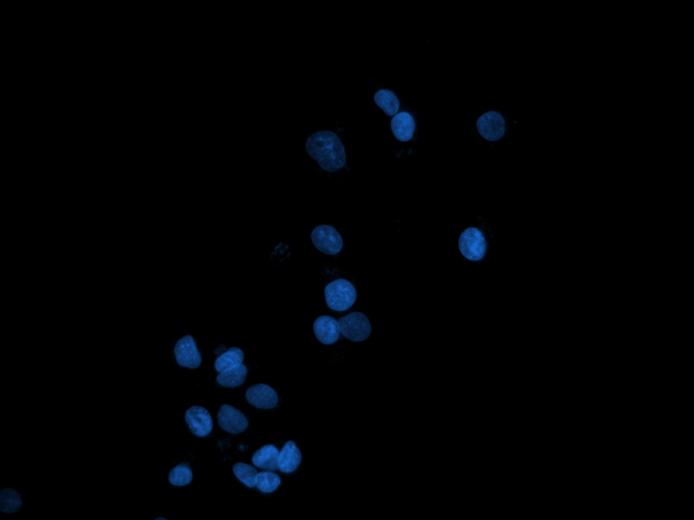
The cell death is one of the essential processes in living organisms. Cells could die as a result of regular and programmed processes (e.g., apoptosis) as well as they die due to premature and unnatural events such as trauma and inflammation (e.g., necrosis). It is thought that the ratio between the dead and live cells gives important information about a number of diseases. For instance, apoptotic index, which is computed in this way, help identify the cancer type and its stage and understand the response to the treatment. Up to date, several biological studies focus on the identification of the relation between diseases and apoptosis/necrosis. Besides these, more recent studies show that diseases may also correlate with other cell death types such as autophagy (digestion of a cell by its own enzymes) and senescence (death as a result of a cell being aged). Understanding the relation between diseases and cell deaths constitutes an important step towards understanding the diseases and drug development against them.
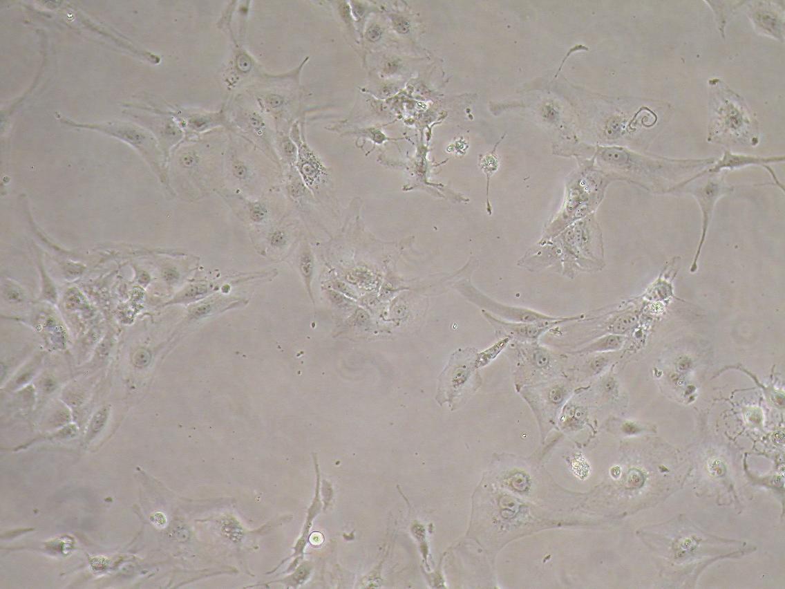
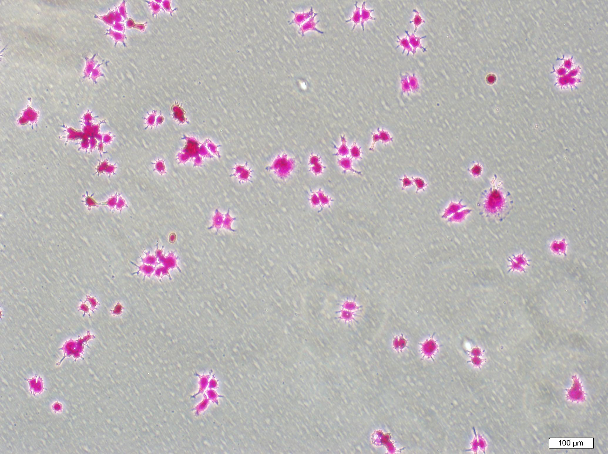
In anticancer drug discovery, a method that is developed by NCI (US National Cancer Institute) is commonly used for measuring the effectiveness of the drug. This method measures the cell response to the drug in terms of whether or not the drug kills cells; here the cell death type and its count are not specified (this test provides indirect information by total protein measurement). However, the classification and quantification of the cell death is very important for drug development. For this purpose, the classification and quantification are commonly achieved by counting the dead and live cells under a microscope. For in vitro bioactivity screening, in which hundreds of chemicals are examined, the counting process that will be done for each drug could be very time consuming, subjective, and irreproducible. Additionally, the NCI test could result in false positives for the cells that are in the process of a cell death but that do not complete this process; such cells could only be observed under a microscope. Therefore, it is very important to develop an image-based screening test for the automatic and objective classification and quantification of cell deaths.
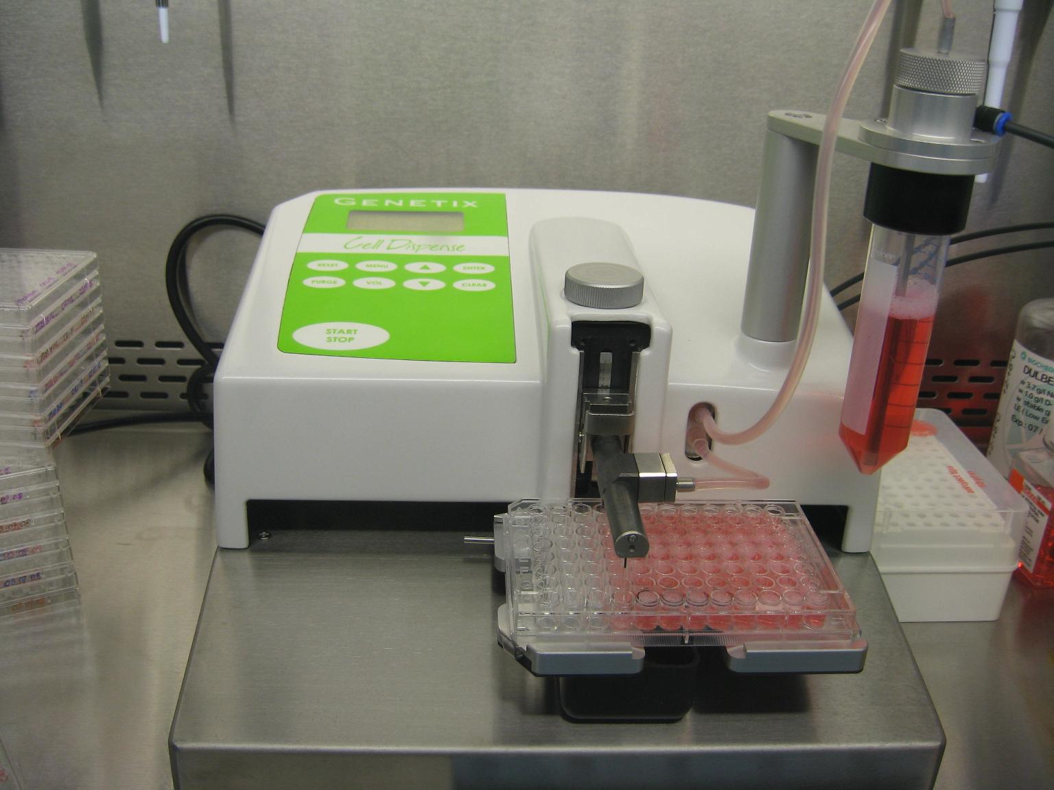
The aim of this project is to develop an objective and intelligent system that expresses the response of live cells to anticancer drug candidates in terms of cell death types and their quantities. This system intends to automatically analyze microscopic images and to automatically classify and quantify cell deaths under a microscope. The proposed project has five main steps. 1.) Growing live cell cultures in media that contain anticancer chemicals and acquiring their microscopic images, 2.) segmenting cells for the identification of their locations, 3.) extracting mathematical features from the segmented cells, 4.) classifying the cells as live or death (apoptotic, autophagic, or senescent) using these features and quantifying their indices, and 5.) developing a new anticancer drug screening test that is based on microscopic imaging and that uses the cell death information.
 |
 |
 |
 |